

Thanh bên bài viết
Số lượt xem pdf: 42
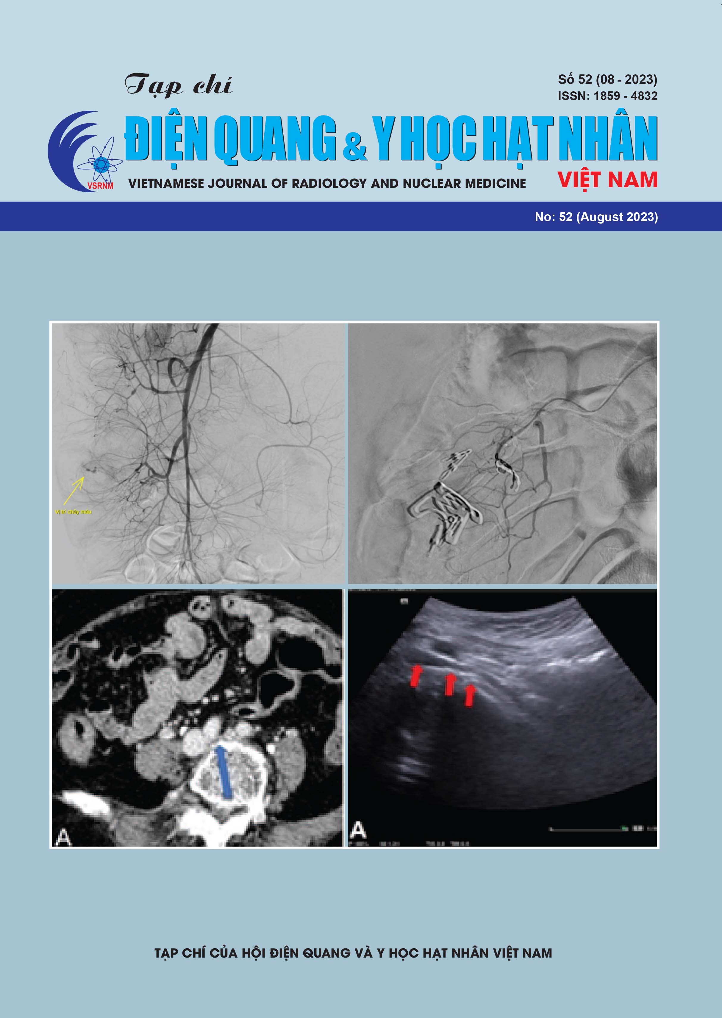
VAI TRÒ CỦA FDG PET/CT TRONG CHẨN ĐOÁN VÀ TIÊN LƯỢNG U PHYLLODES TUYẾN VÚ DI CĂN VÀ HỒI CỨU Y VĂN
Nội dung chính của bài viết
Tóm tắt
U phyllodes ác của tuyến vú tái phát di căn là một căn bệnh rất hiếm gặp với chỉ số ít trường hợp được báo cáo trong y văn. Nhằm cung cấp thêm dữ liệu và hướng dẫn chẩn đoán, cũng như bàn thêm về điều trị và tiên lượng, chúng tôi trình bày một số thông tin về sử dụng chụp PET/CT cũng như phương pháp chẩn đoán hình ảnh khác. Báo cáo nhân một case lâm sàng bệnh nhân nữ, 43 tuổi, được chẩn đoán U phyllodes độ II đã phẫu thuật và xạ trị bổ trợ (2017). Theo dõi bằng hình ảnh học sau 6 năm (02/2023) bệnh nhân được chẩn đoán di căn phổi 2 bên và đã phẫu thuật cắt thùy trên phổi trái sau đó. Kết quả chụp chụp PET/CT tại Bệnh viện Chợ Rẫy cho thấy các tổn thương tăng hoạt động chuyển hóa tại phổi, thành ngực, xương ức, tụy, dạ dày, xương đa ổ khả năng di căn từ u phyllodes trước đây. Các phương pháp điều trị U Phyllodes tái phát di căn hiện nay chưa cho thấy hiệu quả và tiên lượng xấu. Tuy nhiên một số hướng đi mới về các thuốc nhắm trúng đích cho thấy hy vọng kéo dài thời gian sống còn và cải thiện chất lượng cuộc sống của bệnh nhân
Chi tiết bài viết
Tài liệu tham khảo
2. Burga AM, Tavassoli FA. Periductal stromal tumor: a rare lesion with low-grade sarcomatous behavior. Am J Surg Pathol. 2003 Mar;27(3):343-8.
3. Faridi SH, Siddiqui B, Ahmad SS, Aslam M. Progression of Fibroadenoma to Malignant Phyllodes Tumour in a 14Year Female. J Coll Physicians Surg Pak. 2018 Jan;28(1):69-71.
4. Ganesh V, el at. Palliative treatment of metastatic phyllodes tumors: a case series. AME Case Rep. 2017 Dec 21;1:9. doi: 10.21037/acr.2017.12.01. PMID: 30263996; PMCID: PMC6155564.
5. Goto W, Kashiwagi S, Takada K, Asano Y, Morisaki T, Noda S, Takashima T, Onoda N, Hirakawa K, Ohira M. [aA Case of a Malignant Phyllodes Tumor That Was Difficult to Distinguish from Stromal Sarcoma]. Gan To Kagaku Ryoho. 2018 Dec;45(13):2429-2431.
6. Guerrero MA, Ballard BR, Grau AMJ So. Malignant phyllodes tumor of the breast: review of the literature and case report of stromal overgrowth.
7. Inyoung Youn, et al, Phyllodes Tumors of the Breast: Ultrasonographic Findings and Diagnostic Performance of Ultrasound-Guided Core Needle Biopsy, Ultrasound in Medicine & Biology, Volume 39, Issue 6, 2013,Pages 987992, ISSN 0301-5629.
8. Kalambo M, Adrada BE, Adeyefa MM, Krishnamurthy S, Hess K, Carkaci S, Whitman GJ. Phyllodes Tumor of the Breast: Ultrasound-Pathology Correlation. AJR Am J Roentgenol. 2018 Apr;210(4):W173-W179.
9. Khangembam BC, et alMalignant Phyllodes Tumor of the Breast Metastasizing to the Vulva: (18)F-FDG CHỤP PET/CT Demonstrating Rare Metastasis from a Rare Tumor. Nucl Med Mol Imaging. 2012 Sep;46(3):232-3. doi: 10.1007/s13139-012-0154-8. Epub 2012 Jul 19. PMID: 24900068; PMCID:PMC4043033.
10. Li T, Li Y, Yang Y, Li J, Hu Z, Wang L, Pu W, Wei T, Lu M. Logistic regression analysis of ultrasound findings in predicting the malignant and benign phyllodes tumor of breast. PLoS One. 2022 Mar 24;17(3):e0265952.
11. Liberman L, Bonaccio E, Hamele-Bena D, Abramson AF, Cohen MA, Dershaw DD. Benign and malignant phyllodes tumors: mammographic and sonographic findings. Radiology. 1996 Jan;198(1):121-4.
12. Ma W, Guo X, et. al. Magnetic resonance imaging semantic and quantitative features analyses: an additional diagnostic tool for breast phyllodes tumors. Am J Transl Res. 2020 May 15;12(5):2083-2092.
13. Mariapaola Cucinotta, et al “A Strange Case of Phyllodes Tumor Detected Using 18F-FDG PET/CT in an Adolescent Patient Affected by Hodgkin Lymphoma: A Possible Pitfall” Clinical Lymphoma Myeloma and Leukemia, Volume 14, Issue 6,2014.
14. Moon, So Hyang MD;et al. Complete remission of giant malignant phyllodes tumor with lung metastasis: A case report. Medicine 98(22):p e15762, May 2019. | DOI: 10.1097/MD.0000000000015762
15. Nguyễn Ngọc Tuấn Anh – Bài giảng Phyllodes Tumor – Khoa CĐHA - Bệnh viện K Trung Ương.
16. Orguc, S., Mavili, S., Açar, Ç.R. et al. Contrast-enhanced spectral mammographic findings of phyllodes tumor of the breast. Egypt J Radiol Nucl Med 53, 110 (2022).
17. Parneet Singh, et al “Rare case of phyllodes tumour of breast with cardiac and pancreatic metastases findings on FDG CHỤP PET/CT”.Journal of Nuclear Medicine Technology February 2023, jnmt.122.265212
18. Ramakant P, et al. Metastatic Malignant Phyllodes Tumor of the Breast: An Aggressive Disease-Analysis of 7 Cases. Indian J Surg Oncol. 2015 Dec;6(4):363-9. doi: 10.1007/s13193-015-0397-9. Epub 2015 Mar 18. PMID: 27065662; PMCID: PMC4809841
19. Tan PH, Thike AA, Tan WJ, Thu MM, Busmanis I, Li H, Chay WY, Tan MH., Phyllodes Tumour Network Singapore. Predicting clinical behaviour of breast phyllodes tumours: a nomogram based on histological criteria and surgical margins. J Clin Pathol. 2012 Jan;65(1):69-76.
20. Tse GMK, Cheung HS, Pang LM, et al. Characterization of Lesions of the Breast with Proton MR Spectroscopy: Comparison of Carcinomas, Benign Lesions, and Phyllodes Tumors. AJR Am J Roentgenol. 2003;181(5):1267– 1272.
21. Wang H, Xue Y, Xu H. 18 F-FDG PET/CT findings in a giant malignant phyllodes breast tumor. Breast J. 2021 Feb;27(2):183-184. doi: 10.1111/tbj.14148. Epub 2020 Dec 27. PMID: 33368827.
22. Wang, X., Xie, L., Hu, W. et al. Apatinib treatment is effective for metastatic malignant phyllodes tumors of the breast: a case report. BMC Women’s Health 21, 218 (2021)
23. World Health Organization. Histological typing of breast tumors. Tumori 1982;68:181–98.
24. Y. Wang, Y. Zhang, G. Chen, F. Liu, C. Liu, T. Xu, et al. Huge borderline phyllodes breast tumor with repeated recurrences and progression toward more malignant phenotype: a case report and literature review
25. Yasir S, Nassar A, Jimenez RE, Jenkins SM, Hartmann LC, Degnim AC, Frost M, Visscher DW. Cellular fibroepithelial lesions of the breast: A long term follow up study. Ann Diagn Pathol. 2018 Aug;35:85-91.
26. Yoo JL, Woo OH, Kim YK, Cho KR, Yong HS, Seo BK, Kim A, Kang EY. Can MR Imaging contribute in characterizing well-circumscribed breast carcinomas? Radiographics. 2010 Oct;30(6):1689-702.
27. Zhou ZR, Wang CC, Yang ZZ, Yu XL, Guo XM. Phyllodes tumors of the breast: diagnosis, treatment and prognostic factors related to recurrence. J Thorac Dis. 2016 Nov;8(11):3361-3368. doi: 10.21037/jtd.2016.11.03. PMID: 28066617; PMCID: PMC5179374